Iron deficiency anemia (IDA) is the most common form of anemia worldwide and remains a significant health concern in tropical countries where parasitic infections are endemic. Hookworm infestation is a leading but frequently overlooked etiology due to its subtle clinical manifestations and limited routine parasitological testing. We describe two contrasting cases of severe hookworm-related IDA from North Sulawesi, Indonesia: a 72-year-old woman and a 1-year-old infant. Both patients presented with profound microcytic hypochromic anemia but without overt gastrointestinal bleeding. Laboratory evaluation confirmed severe iron deficiency, while stool microscopy revealed hookworm eggs at early cleavage stages, establishing the diagnosis. Despite similar causes, their clinical courses diverged. The infant, whose anemia was compounded by inadequate weaning nutrition, responded rapidly to transfusion, iron supplementation, and single-dose albendazole. The elderly patient, with chronic cumulative exposure from barefoot walking in rural areas, improved more slowly following transfusion, prolonged iron therapy, and a short course of albendazole. These cases highlight the need for routine stool examination in all patients with unexplained IDA in endemic areas, regardless of age. They further emphasize gaps in deworming programs, which often exclude high-risk groups such as infants and older adults. Expanding preventive measures is essential to reduce morbidity and long-term complications.
| Published in | International Journal of Infectious Diseases and Therapy (Volume 10, Issue 4) |
| DOI | 10.11648/j.ijidt.20251004.12 |
| Page(s) | 85-92 |
| Creative Commons |
This is an Open Access article, distributed under the terms of the Creative Commons Attribution 4.0 International License (http://creativecommons.org/licenses/by/4.0/), which permits unrestricted use, distribution and reproduction in any medium or format, provided the original work is properly cited. |
| Copyright |
Copyright © The Author(s), 2025. Published by Science Publishing Group |
Iron Deficiency Anemia, Hookworm Infection, Indonesia
Parameter | Initial Result | Follow-up Result | Reference Range (female adult) | Unit |
|---|---|---|---|---|
Hemoglobin | 4.3 | 7.0 | 12-16 | g/dL |
Red Blood Cells (RBC) | 2.24 | 3.1 | 4.0-5.2 | ×10⁶/μL |
Hematocrit | 15.1 | 23.1 | 36-46 | % |
MCV | 67.9 | 74.5 | 80-96 | fL |
MCH | 19.2 | 22.6 | 27-33 | pg |
MCHC | 28.5 | 30.3 | 32-36 | g/dL |
RDW | 18.0 | 19.9 | 11.5-14.5 | % |
White Blood Cells (WBC) | 8.6 | 3.7 | 4.0-11.0 | ×10³/μL |
Neutrophils | 86 | - | 50-70 | % |
Lymphocytes | 8 | - | 20-40 | % |
Monocytes | 5 | - | 2-8 | % |
Eosinophils | 1 | - | 1-5 | % |
Basophils | 0 | - | 0-1 | % |
Platelets (PLT) | 161 | 218 | 150-400 | ×103/μL |
Serum Iron | 28 | - | 60-170 | µg/dL |
TIBC | 282 | - | 250-450 | µg/dL |
UIBC | 204 | - | - | µg/dL |
Transferrin Saturation | 10 | - | 20-50 | % |
Serum Ferritin | 43.94 | - | 15-150 | ng/mL |
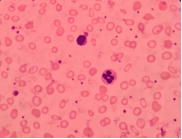
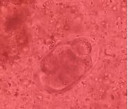
Stool (Ova/Parasite) | Positive (6-cell stage) | - | Negative | - |
Stool RBC/WBC | 5-10 / 2-3 per LPF | - | - | cells/LPF |
Parameter | Initial Result | Follow-up Result | Reference Range (Infant) | Unit |
|---|---|---|---|---|
Hemoglobin | 2.1 | 10.2 | 9.6-13.5 | g/dL |
Red Blood Cells (RBC) | 1.63 | 4.3 | 4.0-5.2 | ×106/μL |
Hematocrit | 9.2 | 34.1 | 34-48 | % |
MCV | 56.4 | 79.3 | 76-92 | fL |
MCH | 12.9 | 23.7 | 23-31 | pg |
MCHC | 22.8 | 29.9 | 32-36 | g/dL |
RDW | 21.0 | 26.7 | 11.5-14.5 | % |
White Blood Cells (WBC) | 10.3 | 12.4 | 5.5-17.5 | ×103/μL |
Neutrophils | 48 | - | 22-46 | % |
Lymphocytes | 36 | - | 37-73 | % |
Monocytes | 10 | - | 2-11 | % |
Eosinophils | 6 | - | 1-4 | % |
Basophils | 0 | - | 0-2 | % |
Platelets (PLT) | 294 | 213 | 150-400 | ×103/μL |
Serum Iron | 37 | - | 40-100 | µg/dL |
TIBC | 491 | - | 250-450 | µg/dL |
UIBC | 454 | - | - | µg/dL |
Transferrin Saturation | 8 | - | 20-50 | % |
Serum Ferritin | 12.18 | - | 20-200 | ng/mL |
Stool (Ova/Parasite) | Positive (6-cell stage) | - | Negative | - |
Stool RBC/WBC | 4-5 / 1-2 per LPF | - | - | cells/LPF |
IDA | Iron Deficiency Anemia |
WHO | World Health Organization |
STH | Soil Transmitted Helminths |
MCV | Mean Corpuscular Volume |
MCH | Mean Corpuscular Hemoglobin |
MCHC | Mean Corpuscular Hemoglobin Concentration |
RDW | Red Cell Distribution Width |
TIBC | Total Iron Binding Capacity |
UIBC | Unsaturated iron Binding Capacity |
RBC | Red Blood Cells |
WBC | White Blood Cells |
PLT | Platelet |
CBC | Complete Blood Count |
| [1] | Kumar A, Sharma E, Marley A, Samaan MA, Brookes MJ. Iron deficiency anaemia: pathophysiology, assessment, practical management. BMJ Open Gastroenterol. 2022 Jan; 9(1): e000759. |
| [2] | Manish A. Iron deficiency anemia: A global public health concern. Int J Clin Biochem Res. 2025 Jan 28; 11(4): 229-36. |
| [3] | Kumar SB, Arnipalli SR, Mehta P, Carrau S, Ziouzenkova O. Iron deficiency anemia: efficacy and limitations of nutritional and comprehensive mitigation strategies. Nutrients. 2022 Jul 20; 14(14): 2976. |
| [4] | Caldrer S, Ursini T, Santucci B, Motta L, Angheben A. Soil-transmitted helminths and anaemia: A neglected association outside the tropics. Microorganisms. 2022 May 13; 10(5): 1027. |
| [5] | Riaz M, Aslam N, Zainab R, Aziz-Ur-Rehman, Rasool G, Ullah MI, et al. Prevalence, risk factors, challenges, and the currently available diagnostic tools for the determination of helminths infections in human. Eur J Inflamm. 2020 Jan; 18: 205873922095991. |
| [6] | Sorisi AMH, Sapulete IM, Pijoh VD. Prevalensi infeksi cacing usus soil transmitted helminths pada orang dewasa di Sulawesi Utara. JKKT. 2019; 7(2):281-4. |
| [7] | Wei KY, Yan Q, Tang B, Yang SM, Zhang PB, Deng MM, et al. Hookworm infection: A neglected cause of overt obscure gastrointestinal bleeding. Korean J Parasitol. 2017 Aug 31; 55(4): 391-8. |
| [8] | Chaparro CM, Suchdev PS. Anemia epidemiology, pathophysiology, and etiology in low‐ and middle‐income countries. Ann N Y Acad Sci. 2019 Aug; 1450(1): 15-31. |
| [9] | Giraldo-Forero JC, Muñoz-Niño LA, Coronado-Castiblanco KV. Severe iron anemia deficiency caused by hookworm: Case report and literature review. Rev Medica Hosp Gen Mexico. 2019 Oct 29; 82(4): 3102. |
| [10] | Lapat JJ, Opee J, Apio MC, Akello S, Ojul CL, Onekalit R, et al. A One health approach toward the control and elimination of soil-transmitted helminthic infections in endemic areas. IJID One Health. 2024 Mar; 2: 100021. |
| [11] | Fairweather-Tait SJ, Wawer AA, Gillings R, Jennings A, Myint PK. Iron status in the elderly. Mech Ageing Dev. 2014 Mar; 136-137: 22-8. |
| [12] | Ahmad A, Madanijah S, Dwiriani CM, Kolopaking R. Iron deficiency risk factors in undernourished children aged 6-23 months in Aceh, Indonesia. J Gizi Dan Pangan. 2018 Nov 15; 13(3): 144-56. |
| [13] | Brooker S, Hotez PJ, Bundy DAP. Hookworm-related anaemia among pregnant women: A systematic review. Raso G, editor. PLoS Negl Trop Dis. 2008 Sep; 2(9): e291. |
| [14] | Abah A, Wokem G. Evaluation of hookworm infections and some haematological parameters amongst primary and secondary schools children in Aba Metropolis, Abia State, Nigeria. J Appl Sci Environ Manag. 2017 Feb 1; 20(4): 935. |
| [15] | Tiremo S, Shibeshi M. Endoscopic diagnosis of hookworm disease in a patient with severe iron deficiency anemia: A case report. Int Med Case Rep J. 2023 Dec; 16: 841-5. |
| [16] | Gaisinskaya P, Sugerik S, Gebara CM. Ascaris lumbricoides diagnosed during evaluation of iron deficiency anemia by capsule endoscopy. Cureus. 2022 May; 14(5): e25208. |
| [17] | Leder K, Weller PF. Eosinophilia and helminthic infections. Best Pract Res Clin Haematol. 2000 Jun; 13(2): 301-17. |
| [18] | Gaskell H, Derry S, Andrew Moore R, McQuay HJ. Prevalence of anaemia in older persons: systematic review. BMC Geriatr. 2008 Dec; 8(1): 1. |
| [19] | Lozoff B, Beard J, Connor J, Felt B, Georgieff M, Schallert T. Long-lasting neural and behavioral effects of iron deficiency in infancy. Nutr Rev. 2008 Jun 28; 64: S34-43. |
| [20] | Diana M, Rawanita M, Natasha CA. Mass deworming program in Indonesia as a strategic tool to reduce soil-transmitted helminth (sth) burden in high-risk population: A systematic review. JKS. 2024; 24(2): 37454. |
| [21] | Naing C, Tung WS, Htet NH, Aung HH, Whittaker MA. Community engagement in health services research on soil-transmitted helminthiasis in Asia Pacific region: Systematic review. Standley CJ, editor. PLOS Glob Public Health. 2023 Mar 22; 3(3): e0001694. |
| [22] | Randell M, Li M, Rachmi CN, Jusril H, Abimbola S, Tama AY, et al. ‘Of’ the community but not ‘of’ the health system: Translating community health workers’ knowledge into credible advice in Aceh, Indonesia. Discov Health Syst. 2024 Feb 1; 3(1): 5. |
| [23] | Albonico M, Montresor A, Crompton DWT, Savioli L. Intervention for the control of soil-transmitted helminthiasis in the community. Adv Parasitol. 2006 Jun; 61: 311-48. |
| [24] | WHO. Preventive chemotherapy in human helminthiasis: coordinated use of anthelminthic drugs in control interventions: a manual for health professionals and programme managers. World Health Organization. 2006; 61. |
| [25] | Ness TE, Agrawal V, Bedard K, Ouellette L, Erickson TA, Hotez P, et al. Maternal hookworm infection and its effects on maternal health: a systematic review and meta-analysis. Am J Trop Med Hyg. 2020 Nov; 103(5): 1958-68. |
| [26] | Vaz Nery S, Pickering AJ, Abate E, Asmare A, Barrett L, Benjamin-Chung J, et al. The role of water, sanitation and hygiene interventions in reducing soil-transmitted helminths: interpreting the evidence and identifying next steps. Parasit Vectors. 2019 Dec; 12(1): 273. |
APA Style
Purwanto, D. S., Tuda, J. S. B. (2025). Hookworm - Associated Iron Deficiency Anemia at Age Extremes: Two Case Reports from North Sulawesi, Indonesia. International Journal of Infectious Diseases and Therapy, 10(4), 85-92. https://doi.org/10.11648/j.ijidt.20251004.12
ACS Style
Purwanto, D. S.; Tuda, J. S. B. Hookworm - Associated Iron Deficiency Anemia at Age Extremes: Two Case Reports from North Sulawesi, Indonesia. Int. J. Infect. Dis. Ther. 2025, 10(4), 85-92. doi: 10.11648/j.ijidt.20251004.12
AMA Style
Purwanto DS, Tuda JSB. Hookworm - Associated Iron Deficiency Anemia at Age Extremes: Two Case Reports from North Sulawesi, Indonesia. Int J Infect Dis Ther. 2025;10(4):85-92. doi: 10.11648/j.ijidt.20251004.12
@article{10.11648/j.ijidt.20251004.12,
  author = {Diana Shintawati Purwanto and Josef Sem Berth Tuda},
  title = {Hookworm - Associated Iron Deficiency Anemia at Age Extremes: Two Case Reports from North Sulawesi, Indonesia},
  journal = {International Journal of Infectious Diseases and Therapy},
  volume = {10},
  number = {4},
  pages = {85-92},
  doi = {10.11648/j.ijidt.20251004.12},
  url = {https://doi.org/10.11648/j.ijidt.20251004.12},
  eprint = {https://article.sciencepublishinggroup.com/pdf/10.11648.j.ijidt.20251004.12},
  abstract = {Iron deficiency anemia (IDA) is the most common form of anemia worldwide and remains a significant health concern in tropical countries where parasitic infections are endemic. Hookworm infestation is a leading but frequently overlooked etiology due to its subtle clinical manifestations and limited routine parasitological testing. We describe two contrasting cases of severe hookworm-related IDA from North Sulawesi, Indonesia: a 72-year-old woman and a 1-year-old infant. Both patients presented with profound microcytic hypochromic anemia but without overt gastrointestinal bleeding. Laboratory evaluation confirmed severe iron deficiency, while stool microscopy revealed hookworm eggs at early cleavage stages, establishing the diagnosis. Despite similar causes, their clinical courses diverged. The infant, whose anemia was compounded by inadequate weaning nutrition, responded rapidly to transfusion, iron supplementation, and single-dose albendazole. The elderly patient, with chronic cumulative exposure from barefoot walking in rural areas, improved more slowly following transfusion, prolonged iron therapy, and a short course of albendazole. These cases highlight the need for routine stool examination in all patients with unexplained IDA in endemic areas, regardless of age. They further emphasize gaps in deworming programs, which often exclude high-risk groups such as infants and older adults. Expanding preventive measures is essential to reduce morbidity and long-term complications.},
 year = {2025}
}
TY  - JOUR
T1  - Hookworm - Associated Iron Deficiency Anemia at Age Extremes: Two Case Reports from North Sulawesi, Indonesia
AU  - Diana Shintawati Purwanto
AU  - Josef Sem Berth Tuda
Y1  - 2025/12/19
PY  - 2025
N1  - https://doi.org/10.11648/j.ijidt.20251004.12
DO  - 10.11648/j.ijidt.20251004.12
T2  - International Journal of Infectious Diseases and Therapy
JF  - International Journal of Infectious Diseases and Therapy
JO  - International Journal of Infectious Diseases and Therapy
SP  - 85
EP  - 92
PB  - Science Publishing Group
SN  - 2578-966X
UR  - https://doi.org/10.11648/j.ijidt.20251004.12
AB  - Iron deficiency anemia (IDA) is the most common form of anemia worldwide and remains a significant health concern in tropical countries where parasitic infections are endemic. Hookworm infestation is a leading but frequently overlooked etiology due to its subtle clinical manifestations and limited routine parasitological testing. We describe two contrasting cases of severe hookworm-related IDA from North Sulawesi, Indonesia: a 72-year-old woman and a 1-year-old infant. Both patients presented with profound microcytic hypochromic anemia but without overt gastrointestinal bleeding. Laboratory evaluation confirmed severe iron deficiency, while stool microscopy revealed hookworm eggs at early cleavage stages, establishing the diagnosis. Despite similar causes, their clinical courses diverged. The infant, whose anemia was compounded by inadequate weaning nutrition, responded rapidly to transfusion, iron supplementation, and single-dose albendazole. The elderly patient, with chronic cumulative exposure from barefoot walking in rural areas, improved more slowly following transfusion, prolonged iron therapy, and a short course of albendazole. These cases highlight the need for routine stool examination in all patients with unexplained IDA in endemic areas, regardless of age. They further emphasize gaps in deworming programs, which often exclude high-risk groups such as infants and older adults. Expanding preventive measures is essential to reduce morbidity and long-term complications.
VL  - 10
IS  - 4
ER  -
Department of Biochemistry, Faculty of Medicine, Sam Ratulangi University, Manado, Indonesia;Department of Clinical Laboratory, Kandou General Hospital, Manado, Indonesia
Biography: Diana Shintawati Purwanto is an associate professor at Sam Ratulangi University, Biochemistry Department. She completed her Master of Laboratory Medicine from The School of Health and Biomedical Sciences at Royal Melbourne Institute of Technology, Australia in 2009, and her Clinical Pathologist Specialization Doctor from Universitas Indonesia in 2018. Apart from being a lecturer, she also works in several hospitals including Department of Clinical Laboratory of Kandou Central General Hospital, Awaloei Hospital, and Budi Mulia Bitung Hospital, North Sulawesi Indonesia, as a clinical pathology specialist. So far, she has been involved in studies related to communicable and non-communicable diseases in North Sulawesi, Indonesia. She also has participated in multiple international research collaboration projects in recent years, especially with Indonesia National Research and Innovation Agency.
Research Fields: hematology, clinical chemistry, infectious disease, biomolecular, entomology.

Department of Parasitology, Faculty of Medicine, Sam Ratulangi University, Manado, Indonesia